Osseous Stracture Human Body : Musculoskeletal System Anatomy And Functions Kenhub - Bone anatomy of upper extremity 12 photos of the bone anatomy of upper extremity anatomy of upper limb bones pdf, bone anatomy of the upper limb, bone anatomy of upper extremity, bone anatomy of upper limb ppt, skeletal anatomy of the upper extremity, bone.
Osseous Stracture Human Body : Musculoskeletal System Anatomy And Functions Kenhub - Bone anatomy of upper extremity 12 photos of the bone anatomy of upper extremity anatomy of upper limb bones pdf, bone anatomy of the upper limb, bone anatomy of upper extremity, bone anatomy of upper limb ppt, skeletal anatomy of the upper extremity, bone.. It contains about 206 bones of different sizes and shapes. Skeletal system | learn skeletal anatomy / in addition to supporting the body's structure against the force of gravity and allowing locomotion, the skeleton plays the vital role of protecting it consists of osseous tissue made of osteocytes, or bone cells. It consists of two types of bone; Human body, the physical substance of the human organism. Human body anatomy is a branch of biology that deals with the study of the structure of the human body.
The human skeleton is the internal framework of the human body. Cartilage helps give structure to the larynx and nose. As part of the body, the spine is a unique and complex structure. It is weaker and easier to fracture than cortical bone, which makes up the shafts of long bones. The term 'anatomy' derives from ancient greek meaning 'dissection' or 'to dissect' and involves the study of the structure of the human body.
Thoracic Spine from www.spineuniverse.com It consists of two types of bone; As part of the body, the spine is a unique and complex structure. Tremendous advantages have been gained from this erect posture, the chief among which has been the freeing of the arms for a great variety of uses. Cartilage helps give structure to the larynx and nose. The positioning and articulation of the bones of the skeleton also determine the nature of the body's movements. This science quiz game will help you learn 15 of the most important bones. A system is an organization of varying numbers and kinds of organs so arranged that together they can perform. Visualized osseous structures that are unremarkable in a chest xray means that.
The diaphysis and the epiphysis.the diaphysis is the tubular shaft that runs between the proximal and distal ends of the bone.
Tremendous advantages have been gained from this erect posture, the chief among which has been the freeing of the arms for a great variety of uses. A distinctive characteristic of humans as compared with other mammals is erect posture. Learn about the main tissue types and organ systems of the body and how they work together. These bones provide structure and protection and facilitate motion. Learn more about the composition, form, and physical adaptations of the human body. It consists of two types of bone; Human bones diagram 12 photos of the human bones diagram human anatomy diagram back view organs, human anatomy diagram diaphragm, human anatomy diagram of ear, human anatomy torso diagram, human skeleton diagram with labels, bone, human anatomy diagram back view organs, human anatomy diagram diaphragm, human anatomy diagram of ear, human. Cartilage helps give structure to the larynx and nose. Skeletal system | learn skeletal anatomy / in addition to supporting the body's structure against the force of gravity and allowing locomotion, the skeleton plays the vital role of protecting it consists of osseous tissue made of osteocytes, or bone cells. It is composed of 300 bones at birth, but later decreases to 80 bones in the axial skeleton and 126 bones in the appendicular skeleton. The largest bone in the human body is the thighbone or femur, and the smallest is the stapes in the middle ear, which are just 3 millimeters (mm) long. It's the bone in your leg that goes from your hip to your knee. It contains about 206 bones of different sizes and shapes.
Cartilage helps give structure to the larynx and nose. Human body anatomy is a branch of biology that deals with the study of the structure of the human body. It's the bone in your leg that goes from your hip to your knee. Learn about the main tissue types and organ systems of the body and how they work together. The femur forms the ball and socket hip joint with the hip bone and forms the knee joint with the tibia and patella.
Glossary Of The Skeletal System Learn Skeletal Anatomy from www.visiblebody.com Related posts of bone structure of human body in 3d bone anatomy of upper extremity. It is composed of many different types of cells that together create tissues and subsequently organ systems. Osseous stracture human body : In an average adult, it's about 20 inches long. Some, like the rib cage, provide protection for softer body parts, while other bones enable mobility by supporting the muscles. The human body is made up of 11 organ systems that work with one another (interdependantly). The longest bone in the human is called the femur, or thigh bone. Visualized osseous structures that are unremarkable in a chest xray means that.
The term 'anatomy' derives from ancient greek meaning 'dissection' or 'to dissect' and involves the study of the structure of the human body.
It's the bone in your leg that goes from your hip to your knee. The bone mass in the skeleton reaches maximum density around age 21. This includes the head, facial, hyoid, auditory, trunk, ribs, and sternum. Cartilage is more flexible than bone but stiffer than muscle. Bones are organs that produce red and white blood cells, store minerals, enable mobility, and provide structural support for the body.they are lightweight, strong, and hard, and function within the body in many different processes, including autoimmune function. It contains about 206 bones of different sizes and shapes. Some, like the rib cage, provide protection for softer body parts, while other bones enable mobility by supporting the muscles. Bones have an internal structure similar. 1 iv knee anatomy i. The structure of a long bone allows for the best visualization of all of the parts of a bone ().a long bone has two parts: The collection of bones in the human body is called the skeletal system. The human body is the structure of a human being. The human body is made up of several organ systems that work together as one unit.
The human body is to some extent like a walking tower that moves on pillars, represented by the legs. 1 iv knee anatomy i. Classification of bones in the human body. The human skeletal system consists of all of the bones, cartilage, tendons, and ligaments in the body. Storage of calcium, other minerals, and fat.
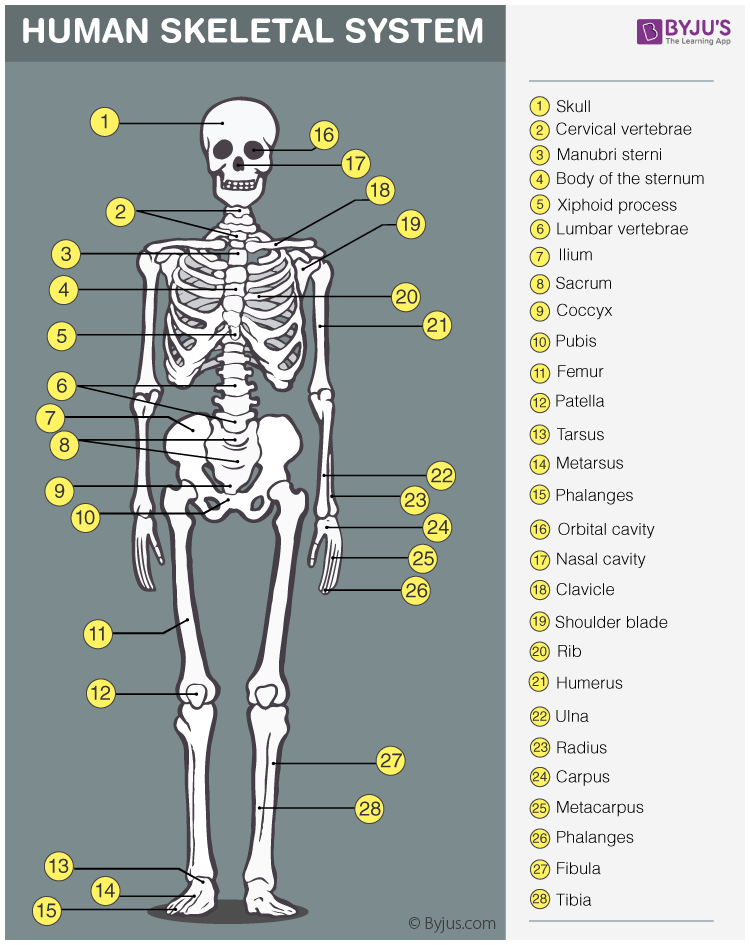
Skeletal System Anatomy Physiology Of Human Skeletal System from cdn1.byjus.com The term 'anatomy' derives from ancient greek meaning 'dissection' or 'to dissect' and involves the study of the structure of the human body. Osseous stracture human body / human anatomy 3d view ~ human anatomy. There are 206 bones in the human skeleton, not including teeth and sesamoid bones (small bones found within cartilage): The human body consists of eleven organ systems , each of which contains several specific organs. Terms relating to anatomical structures and directions. It is composed of 300 bones at birth, but later decreases to 80 bones in the axial skeleton and 126 bones in the appendicular skeleton. Visualized osseous structures that are unremarkable in a chest xray means that. The human body is to some extent like a walking tower that moves on pillars, represented by the legs.
It's only about 3 millimeters long in an adult.
Human body anatomy is a branch of biology that deals with the study of the structure of the human body. The bone mass in the skeleton reaches maximum density around age 21. Human body hair may seem annoying to groom, pluck and shave, but it does. Bone anatomy of upper extremity 12 photos of the bone anatomy of upper extremity anatomy of upper limb bones pdf, bone anatomy of the upper limb, bone anatomy of upper extremity, bone anatomy of upper limb ppt, skeletal anatomy of the upper extremity, bone. Bones are organs that produce red and white blood cells, store minerals, enable mobility, and provide structural support for the body.they are lightweight, strong, and hard, and function within the body in many different processes, including autoimmune function. The structure of a long bone allows for the best visualization of all of the parts of a bone ().a long bone has two parts: 1 iv knee anatomy i. The femur forms the ball and socket hip joint with the hip bone and forms the knee joint with the tibia and patella. The femur is the largest bone in the body and the only bone of the thigh (femoral) region. It is very porous and contains red bone marrow, where blood cells are made. Terms relating to anatomical structures and directions. The human body is made up of 11 organ systems that work with one another (interdependantly). The term 'anatomy' derives from ancient greek meaning 'dissection' or 'to dissect' and involves the study of the structure of the human body.


0 Komentar